Feline Resorptive Lesions (‘FORLS’)
This common condition affects more than one third of adult cats. The cause remains unclear but the signs and symptoms are evident on clinical exam. The condition results in one or more teeth spontaneously ‘dissolving’ away. The result is severe pain while the tooth undergoes change.
There are two recognised types of FORLS:
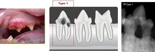
Type 1
These lesions are associated with periodontal disease. They occur at the neck of the tooth, just above the gumline. The gum surrounding the lesion is usually inflamed. On X-ray – the crown (the portion of the tooth seen above the gumline) has undergone resorption but the root (the portion of tooth below the gumline, hidden from the naked eye) has remained intact.
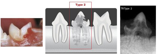
Type 2
These resorptive lesions begin on the root surface. The tooth crown may appear pink with a small rounded portion of bright red ‘gum like’ tissue protruding from it (granulation tissue). On X-ray the root has been turned into the bone (ankylosis) and cannot be seen.
Signs of oral pain in cats
Oral pain is often harder to spot in cats than you may imagine. EVEN SEVERE DENTAL PAIN WILL NOT STOP A CAT EATING! In the same way we manage to eat around a sore tooth, our pets do the same.
- Slow eating
- Bleeding gums
- Drooling
- Shows interest in food then backs away
- Choosing to only eat on one side of the mouth
- Pain on allowing oral examination
- Pawing at mouth in severe cases
- Weight loss
A good way to tell if your cat is eating on one side is to check in the mouth. If the teeth on one side of the mouth look ‘cleaner’ than the other side, this is often an indication that the cat is choosing that side to eat on. As the cat chews food, it keeps these teeth clean and therefore the opposite side usually has signs of disease.
Treatment
As evident above, the only way to distinguish the different types of resorptive lesions is with X-ray. This is an important first step as this then determines the treatment. The treatment of choice is to extract any affected tooth as the lesion will only progress and if left in situ could be a permanent source of discomfort for the cat.
Type 1 FORLS – these teeth have their root structure intact and the roots will not dissolve. Eventually the crown fractures off leaving open tooth roots exposed to the mouth. This is extremely painful. To remove the root structure, we will often perform a surgical extraction, which involves cutting the gum, removing the tooth and then suturing the gum back together.
Type 2 FORLS – potentially, if the gum is healthy, these teeth can have the crown of the tooth amputated and the root left to naturally turn into bone. However, if the tooth is affected by other disease processes or the gum is extremely inflamed, they should also be removed similar to type 1 FORLS.